
Watch a bird overhead, turn toward a friend at your side, or exult in a cartwheel, and without knowing it, you are taking advantage of the vestibular portion of your inner ear. Combined with visual cues and information from muscles and joints, our inner ears allow us to maintain balance and a steady field of vision during movements.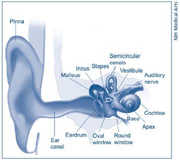
Diagram of the inner ear. (Click on image for larger view.)
A specific part of the inner ear contributes to good balance and steady vision by detecting angular head motions—such as nodding forward, tilting sideways, or looking over your shoulder. It also signals the brain about changes in head position so that simple movements do not leave us feeling imbalanced or give us vertigo.
Scientists are seeking to better understand how our genes direct the development of this portion of the inner ear. Specifically responsible for detecting angular head movements, it is made up of three semicircular canals and their associated sensory tissues, called cristae. The cristae contain sensory cells that detect movement of fluid within the semicircular canals during head movements. The three precisely positioned semicircular canals, which are composed of nonsensory tissue, must develop in an exact geometry—approximately perpendicular to each other and connected to the inner ear's vestibule.
A research team led by Doris Wu, Ph.D., of the National Institute on Deafness and Other Communication Disorders (NIDCD), recently discovered that a gene encoding a particular growth factor protein is essential to the formation of the semicircular canals and the cristae. Dr. Wu and colleagues' earlier research has also identified other genes involved in the well-choreographed cascade of molecular events that form the inner ear.
Working with scientists at NIDCD, Duke University, and the Albert Einstein College of Medicine, Dr. Wu found that bone morphogenetic protein 4, or Bmp4, is required for formation of the semicircular canals and their cristae in mice. When she and her team inactivated or deleted the gene for Bmp4 in the inner ear, the semicircular canals did not develop. Bmp4 has long been known to play an important role in the formation of connective tissues and other organs such as eyes, limbs, and cartilage.
The researchers found that during inner ear development, Bmp4 is secreted by sensory tissue of the cristae in the base of each canal. Based on their new research, they hypothesize that sensory patches in the cristae regulate formation of the nonsensory tissue in the canals. Dr. Wu and her team are seeking a full understanding of the chain of events that give rise to the inner ear.
Dr. Wu and her team's work is published in the April 2008 issue of the journal PLoS Genetics. The paper is available online.
Taken from www.nidcd.nih.gov/
All About Balance: Scientists Identify Building Blocks of Inner Ear Structure
Share: