
Meniere's Disease is a condition that should be understood by all healthcare professionals involved with treating and managing ear disorders, including audiologists and physicians. This article provides an overview of the clinical picture, pathology, etiology, natural history, and medical and surgical treatment of Meniere's Disease.
Clinical Types:
Meniere's Disease can be classified many ways. In our management of Meniere's Disease, we use the following three primary subclassifications of Meniere's Disease, to describe the specific signs and symptoms, as noted below.
- Classic Meniere's Disease is an inner ear disorder characterized by episodic vertigo attacks (often with nausea and vomiting), sensorineural hearing loss, tinnitus, and pressure or fullness in the involved ear (usually unilateral). Initially, the hearing loss typically involves the low frequencies and the hearing loss fluctuates, generally becoming worse with each attack. Over time, the hearing loss progresses to involve the higher frequencies, and the degree of hearing loss can progress to severe-to-profound.
It is estimated that 80 percent of all Meniere's Disease patients are unilaterally involved. - In Vestibular Meniere's Disease, vertigo attacks are identical to classic Meniere's Disease (above). However, in Vestibular Meniere's Disease, hearing remains normal, and other aural symptoms (tinnitus, full-ness) are absent.
- Bilateral Meniere's Disease is characterized by bilateral fluctuating hearing loss and recurrent episodes of vertigo. One ear may initially present and later enter a quiescent period. Years later, disease in the opposite ear may develop. Approximately 50% of patients destined to develop bilateral Meniere's Disease do so within 2 years, and 75% do so within 5 years. If patients with bilateral Meniere's Disease experience symptomatic improvement with oral steroids, a diagnosis of autoimmune inner ear disease is made.
Although Meniere's Disease is a common diagnosis in the otology office, it is not necessarily an easy disorder to diagnose, as many other conditions and clinical presentations must be effectively ruled-out in order to arrive at the diagnosis of Meniere's Disease. For example, vestibular neuronitis, benign positional vertigo, viral labyrinthitis, perilymph fistula, vascular compression of the eighth cranial nerve, chronic labyrinthitis, and circulatory, central nervous system, or metabolic problems can disguise themselves as one of the four sub-classifications discussed above.
Etiology:
Meniere's Disease is likely caused by dilatation (stretching or widening) of the endolymphatic spaces (hydrops) with evidence of ruptures and healing of the membranous labyrinth. It is thought to be due to hypersecre-tion of endolymph by the stria vascularis, or by under absorption of endolymph caused by functional failure of the endolymphatic sac, or a combination of the two processes. However, the exact mechanism for development of hydrops is not entirely understood. Histologic study of temporal bones usually demonstrates a markedly enlarged saccule, with the saccular membrane adherent to the undersurface of the stapes footplate. There is also dilatation of Reissner's membrane in the cochlea and enlargement of the utricle.
Natural History:
It is important to understand the natural history of Meniere's Disease, particularly in order to determine the true impact of the treatment, as compared to the natural course of the disease.
A. Natural History versus Endolymphatic Shunt:
The natural history was studied in a group of 50 control patients with Meniere's Disease who were surgical candidates for endolym-phatic subarachnoid shunt (ELS), but who elected not to undergo surgery.1 The control patients were compared with 89 treated patients who had ELS surgery for Meniere's Disease.
The control group had a 57% spontaneous remission rate at 2 years and a 71% spontaneous remission rate at 8 years. The 89 treated patients who had ELS surgery, had statistically similar control of vertigo spells. In other words, the treated ELS group showed no greater reduction of the attacks of Meniere's Disease than did the control group. It was concluded that the results of endolymphatic sac proce-dures did not alter significantly from the spontaneous remission rate of Meniere's Disease.
B. Natural History versus Vestibular Neurectomy:
Patients who had eighth nerve section or vestibular neurectomy showed statisti-cally greater remission of vertigo after surgery than did the control group that had no surgery. The neurectomy procedures (vestibular neurectomy or eighth nerve section) offer statistically significant improvement of vertigo as compared to the natural history, and they often give patients imme-diate cure of their vertigo.
Incidence of Meniere's Disease:
The incidence of bilateral Meniere's Disease was studied by evaluating 7600 otology charts over a 5-year period.2 Meniere's Disease was found in 3% of the otology charts.
Of 240 patients with Meniere's Disease, 161 were medically treated and 79 were surgically treated. Bilateral Meniere's disease was found in 17% of the medically treated group and in 3.8% of the surgi-cally treated group.
Of the patients who underwent vestibular neurectomy, none developed bilateral Meniere's Disease during the average five year follow-up time period. After cochleovestibular neurectomy, there was a 7% incidence of bilateral Meniere's Disease, and a 9% incidence after endo-lymphatic sac surgery. The reason for the lower incidence of bilateral Meniere's Disease involvement in neurectomy patients as compared with the medically treated patients is not certain. However, by the time most patients have had the disease long enough to require surgery (i.e., 5 years), the second ear statistically will have shown evidence of Meniere's Disease in 75% (as mentioned previously). In this study, 50% of the patients who would develope bilateral Meniere's Disease did so within the first 2 years and 75% developed it by 5 years.
Medical Treatment:
Meniere's Disease has no proven medical cure. Supportive therapy is utilized, and an effort is made to reduce the endolymph volume by prescribing diuretics, such as triamterene-hydrochlorothiazide (Dyazide), and encouraging a low salt diet. When a patient is seen early in the course of the disease, he or she is treated empirically with oral steroids, and the hearing response is measured. If there is a response, which typically occurs in about 10 to 20% of patients, the patient is evaluated for autoimmune inner ear disease. Meclizine, 25 mg three or four times a day as needed, is prescribed for vertigo attacks. However, if the patient is nauseated or vomiting, the drug may be difficult to ingest. In that case, 25 mg prochlorperazine (Compazine) suppositories are used. To reduce the volume of endolymph,the patient is given one tablet of Dyazide a day, on a long-term basis.
For bilateral Meniere's symptoms and signs, autoimmune inner ear disease is suspected. The diagnosis is confirmed if the patient responds to oral steroids. Other treatment options for autoimmune inner ear disease include use of powerful anti-inflammatory chemotherapeutic medications such as methotrexate and inner ear perfusion with steroids.
Surgical Treatment:
Surgical treatment is indicated when a patient is incapacitated with unilateral Meniere's Disease and quality of life is affected. Approximately 20% of patients seen in the physician's office for Meniere's Disease eventually have surgery. Certain criteria for surgery should be met. Hearing should be good in the opposite ear and there should be no ataxia. There must be objective evidence of unilateral inner ear disease, including a sensorineural hearing loss, usually worse in the low frequencies. ENG typically shows a reduced vestibular response in the affected ear in about 50% of cases, and occa-sionally, an elevated summating potential is present on electrocochleography. There must be good balance function and no psychiatric illness or possible secondary gain, such as disability. Surgery is contraindicated in the treatment of Meniere's Disease in an only hearing ear and generally in bilateral disease.
Importantly, the ideal surgical treatment should be minimally invasive, require no more than local anesthesia, reliably induce a complete reduced vestibular response, and preserve hearing with minimal patient morbidity.
Inner Ear Perfusion with Gentamicin:
The least invasive surgical treatment of Meniere's Disease involves gentamicin perfusion of the inner ear. The goal of the procedure is to treat the affected ear with a vestibulotoxic medication to induce a complete vestibular deficit on the treated side while minimizing hearing loss. The advantages of placing medications directly into the inner ear include: 1) the diseased ear is treated directly without affecting the entire body; 2) a higher inner ear concentration of medication can be obtained; and 3) systemic side effects of the drug are prevented.
The authors' technique of choice to perfuse the inner ear involves use of the Silverstein MicroWick™ (Micromedics, Eagan, MN). The Silverstein MicroWick™ is a sponge-like device that allows direct and precise delivery of medication to the round window membrane (Fig. 1).3
Figure 1. Polyvinyl acetate MicroWick™ which measures 1 mm in diameter by 9 mm long. Microwick is manufactured by Micromedics. 1-800-624-5663, www.micromedics-usa.com. This illustration is reprinted with permission from Micromedics. Audiology Online is grateful for Micromedics permission to use this illustration here.
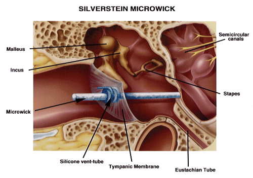
The patient self-administers the medication into the ear canal. The MicroWick™ absorbs the medication and transports it to the round window membrane where it perfuses directly into the inner ear fluids. This method is similar to the current concept of self-trea™ent for eye disease using medicated eye drops.
The essentials of the procedure include anesthetizing the ear with an injected anesthesia, after which a laser tympanostomy or vertical myringotomy is made through the tympanic membrane, overlying the round window niche (see Silverstein, 1999).4 The middle ear is examined with an endoscope or the operating microscope to determine if there are any obstructing membranes over the round window niche. If such membranes are present, a small pick is used to clear them. A ventilation tube is inserted into the tympanostomy and the MicroWick™ is inserted through the tube towards the round window until resistance is met.
Gentamicin is injected into the middle ear and the MicroWick™ is saturated with gentamicin, allowing delivery of a high concentration directly to the round window membrane with resultant perfusion into the inner ear fluids. Patients self-administer medication into the ear three times daily, while lying with the treated ear facing upward for fifteen minutes each time.
At the end of each treatment week, audiometric testing is completed. The weekly audiometric measures include; pure tones by air and bone conduction, word recognition scores, electrocochleography, and balance function by warm and ice-air caloric electronystagmography (ENG). Patients are also questioned regarding changes in their subjective symptoms of vertigo, aural pressure, tinnitus, and imbalance. Depending upon the objective test results and the patient's symptoms, treatment is continued or discontinued.
The goal of the treatment is to obtain a 100% reduced vestibular response to both bithermal and ice-air caloric ENG testing without producing a hearing loss. The usual length of treatment is 2-3 weeks (range 1-6 weeks). During the treatment period, if the hearing significantly decreases but vestibular function is still present, the treatment is discontinued for one week and steroids may be recommended to "rescue" hearing. The patient is re-evaluated one week later, and treatment is reinstituted if the hearing improves. If the vestibular function reaches a 100% reduced vestibular response or remains stable and does not decrease after several weeks of treatment, the treatment is discontinued. At that point, the MicroWick™ and vent tube are removed in the office with a pick, without the need for anesthesia.
Of 92 patients with Meniere's Disease who self-treated using dilute gentamicin, vertigo symptoms were relieved in 85% of the patients responding to a questionnaire.3 Eight percent needed a subsequent surgical procedure for Meniere's Disease. Pressure in the ear was improved or relieved in 67% responding, while tinnitus was relieved or improved in 57% of patients. Thirty-six percent had progression of their hearing loss in the treated ear.
This new technique of self-treatment using the MicroWick™ to deliver medications to the inner ear is a minimally invasive, inexpensive, safe, effective, and well-tolerated method for treating Meniere's Disease.
Vestibular Neurectomy:
If disabling vertigo persists in unilateral Meniere's Disease despite one or more treatments with gentamicin perfusion of the inner ear, other more aggressive surgical alternatives exist. For hearing that is better than 80 dB PTA and has better than 20% word recognition scores, the procedure of choice is a posterior fossa microsurgical vestibular neurectomy, which generally allows hearing preservation (see below).
First described through the retrolabyrinthine approach in 1979, the combined retro-labyrinthine/retrosigmoid vestibular neurectomy is an evolution in our technique and our preferred approach in the posterior fossa.5 In this procedure, after a postauricular skin incision is made, a limited mastoidectomy is performed, and the lateral venous sinus is skeletonized in its course through the mastoid. The posterior fossa is entered through a dural incision made just behind the lateral venous sinus. Retraction sutures are placed in the cuff of dura, allowing forward retraction of the sinus to give excellent exposure to the cranial nerves at the base of the skull. After the spinal fluid is released and the arachnoid is opened, the eighth cranial nerve can be seen traversing the cere-bellopontine angle. This nerve is studied for the presence of a cleavage plane between the cochlear and vestibular nerves, at times seen more clearly near the brainstem and at other times closer to its entry into the internal auditory canal. The surgeon must use high-powered magnification to delineate the separation between the cochlear and vestibular portions of the eighth nerve. The vestibular nerve is usually transected in the posterior fossa. Occasionally, the separation cannot be identified and the posterior lip of the internal audi-tory canal must be drilled down for better exposure of the cleavage plane.
Over 230 vestibular neurectomies have been performed through the authors' practice with excellent results and infrequent complications.6 Ninety-three percent of the patients are free of vertigo and would recommend the procedure to another patient. The dural incision behind the lateral sinus has been used in over 57 cases with no cerebrospinal fluid leak, facial paralysis, or meningitis. One percent of the patients experience total hearing loss and 20% has a postoperative senso-rineural hearing loss worse than 20 dB.
Importantly, most vestibular neurectomy patients have signifi-cant hearing loss before surgery, and very few complain or notice additional postoperative hearing loss, if it occurs. Generally, patients are happy to be free of vertigo attacks. The tinnitus and pressure that may continue do not pose significant problems, and most patients resume a normal life style. Overall, posterior fossa vestibular neurectomy is relatively safe and is a highly effective procedure. The surgical experience throughout the country is similar to ours, indicating a low incidence of complica-tions and a high rate of success (93%) in curing vertigo attacks.
Labyrinthectomy:
When hearing is worse than 80 dB or worse than 20% word recognition score, or when hearing is otherwise not useful, labyrinthectomy with or without transcochlear cochleovestibular neurectomy is recommended.
Both procedures are performed through the ear canal and result in sacrifice of functional hearing. After a typanomeatal flap has been elevated through the ear canal, labyrinthectomy involves drilling of the promontory and opening of the basal turn of the cochlea. Then, the neuroepithelium of the labyrinth is removed with a right angle pick. Due to occasional failure of vertigo control with labyrinthectomy alone, transcochlear cochleovestibular neurectomy was added to the procedure to improve success. This technique allows a fast and the most direct approach to vestibular nerve sectioning. Over 150 procedures have been performed since 1970. This procedure, the gold standard for Meniere's disease surgery, has a high cure rate (88%).7 In almost 70% of patients, this procedure relieves or reduces the tinnitus, pressure, and fullness in the ear. The procedure has proven to be safe, with low incidence of complications, including no cases of facial paralysis.
CONCLUSION:
We know that Meniere's Disease is due to dilation of the endolymphatic spaces, but we are unsure of the exact cause of the hydrops. An accurate diagnosis of Meniere's Disease can be made, primarily based on detailed patient history and audiometric and vestibular testing. The natural history of Meniere's Disease demonstrates a high spontaneous vertigo remission rate over the ensuing years, and all treatments must be compared against this standard.
We can offer patients successful medical and surgical treatment, including innovative new surgical techniques. Meniere's patients no longer need to hear the old adage, "you have to learn to live with it.'' Today, we can offer the patient relief from vertigo. In the future, the cause of Meniere's Disease and a treatment for the associated hearing loss must be found so a true cure can be given to the patient.
REFERENCES:
1. Silverstein H, Smouha E, Jones R. Natural history vs. surgery for Meniere's disease. Otolaryngol Head Neck Surg 1989;100:6-16.
2. Rosenberg S, Silverstein H, Flanzer J, Wanamaker H. Bilateral Meniere's disease in surgical versus nonsurgical patients. Am J Otol 1991;12;336-340.
3. Silverstein H, Lewis WB, Jackson LE, Thompson JH, Rosenberg SI. Self-treatment of inner ear using a MicroWick™. Ear Nose Throat J 2002 (accepted for publication).
4. Silverstein H. The MicroWick™ to deliver medication to the inner ear. Ear Nose Throat J 1999;78:595-600.
5. Silverstein H, Jackson LE. Vestibular nerve section. Otolaryngol Clin North Am 2001 (awaiting publication).
6. Silverstein H, Norrell H, Smouha E, Jones R. Combined retrolab-retrosigmoid vestibular neurectomy. An evolution in approach. Am J Otol 1989;10:166-169.
7. Silverstein H, Jones R, Rosenberg S. Transmeatal cochleovestibular neurectomy. In Operative Techniques in Otolaryngology-Head and Neck Surgery: Surgical Manage-ment of Meniere's Disease. Philadelphia, W.B. Saunders, 1991;2:32-34.
