Tympanometry was first developed in the 1950's by Terkildsen et al to measure middle ear pressure. Over the years, its contribution to clinical diagnosis has become well accepted and it is now a routine part of the audiological test battery.
Tympanograms are used to identify middle ear pathologies. Most commercial tympanometers offer a 226 Hz probe tone. Though it has been shown that other probe frequencies can obtain different results. (Hunter and Margolis, 1992; Marchant et al, 1984; Shurin et al, 1976, 1977).
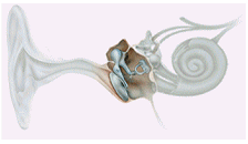
A tympanometer consists of an air pump, a probe with a loud speaker, a microphone and a manometer. The probe tip must be fit snuggly in the ear canal to for a hermetic seal to be formed, which is required to obtain the proper pressure in the ear. The air pump introduces air pressure changes in the ear canal and a probe tone is also sent into the ear canal. The probe mechanism contains a microphone and speaker, which emits a pure tone. The acoustic immittance of the ear is analyzed by continuously monitoring sound pressure levels in the ear canal with a probe microphone. The immittance at the Tympanic membrane (TM) is directly proportional to the sound pressure level of the probe tone in the ear canal.
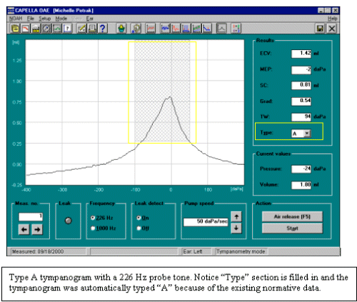
The most commonly used probe tone has been 226Hz. This probe tone has some definitive advantages when testing the adult ear. That's because the adult middle ear system is stiffness-dominated (compliance) at this frequency and the effects of mass and friction are minor. Interpretation and presentation of the results in most instruments display only the compliance of the middle ear. Additionally, the compliance value is directly proportional to the closed air volume, and the external ear canal volume (ECV) is obtained.
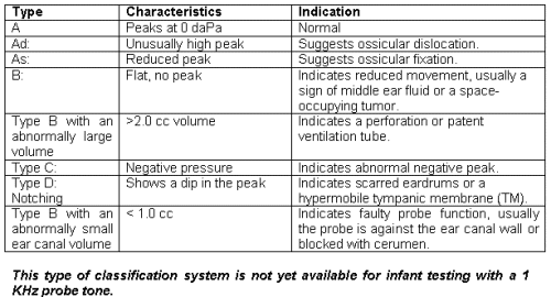
A common classification system for interpreting tympanograms with a 226 Hz probe exists (Linden et al, 1969, modified by Jerger 1970).
Multiple-Frequency Tympanometry
Multiple-frequency tympanometry is a method of measurement that sweeps through a series of frequencies, e.g. from 250 to 2000 Hz. Through multiple-frequency tympanometry it is possible to directly assess the resonant frequency of the middle ear system. The resonant frequency is the probe tone frequency where susceptance becomes zero due to the counteractive forces of its components. A normal value for resonant frequency is around 900Hz. Below this value the middle ear system is stiffness-controlled and above the normal value mass-controlled according to the more prominent component of the susceptance (Shanks et al 1981). The changes in resonant frequency are used to assess the pathology of the middle ear system, especially those of the ossicular chain.
The Infant Ear
The infant ear has a bony region that is not yet completely formed which results in a highly compliant ear canal. During development of the infant ear, several changes take place, which influence the mechanical properties of the ear canal. Early changes that take place include the fusing of the tympanic ring. This process involves mechanical changes, which influence the tympanogram. The external and middle ear systems vary significantly in their acoustic response properties over the first 2 years after birth (Keefe et al, 1993; Keefe and Levi, 1996).
Physical changes in the external and middle ear after birth that could account for the acoustic changes include:
- Size increase of the external ear
- Middle ear cavity and mastoid
- A change in the orientation of the tympanic membrane
- Fusion of the tympanic ring
- A decrease in the overall mass of the middle ear (due to changes in bone density, loss of mesenchyme),
- Tightening of the ossicular joints
- Closer coupling of the stapes to the annular ligament
- The formation of the bony ear canal wall
Tympanometry in Infants:
Tympanograms collected from infant ears progress differently than those collected from adult ears. The infant ear anatomy differs in many ways when compared with the adult ear. Because of these differences, a higher frequency probe tone is needed to collect tympanograms that will be useful in identifying middle ear effusion (MEE). Tympanograms in pediatric audiology are most commonly used to measure middle ear pressure, which assists in the identification of middle ear fluid commonly referred to as otitis media. 1KHz probe tones are available in a number of tympanometers today. Tympanometry is commonly included in the diagnostic test when ABR or OAE are abnormal during follow-up testing after infants refer from an ABR or OAE screening protocol.
Earlier research shows infants below 4 months may demonstrate what appears to be a normal 220Hz tympanogram even with confirmed MEE. (Paradise et al 1976, Shurin et al 1976, Meyer et al 1997). It is also possible to obtain what appears to be abnormal 220Hz tympanograms in normal ears (Keefe et al 1996). It was originally believed that the baby's ear canal walls were so compliant that a movement of the entire canal wall occurred that imitated the type A tympanogram (Holte et al., 1991). More recently it is thought that there may be a better correlation between the presence of middle ear effusion and the shape of the tympanogram when a high-frequency probe tone is used. (Marchant et al., 1984). Some researchers have concluded that higher probe tone frequency tympanometry can accurately identify MEE. (Shurin et al 1977, Marchant et al 1984)
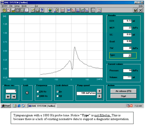
At this time, as further research continues to support high frequency tympanometry, the best choice for a tympanometric probe frequency in babies under 4 months of age is 1000 Hz. Research facilities are currently collecting data on normative values. But as of this time, there are still many unknowns regarding sensitivity and specificity of 1000-Hz tympanometry to the presence of middle ear effusion in infants. And to date, there is not a normative data classification system in place, such as we have for the 226 Hz probe tone in adults. So, it is recommend that high frequency tympanometry be included in a battery of tests to identify the abnormality present in an infant, but caution be taken in the interpretation. Tympanometry is most effective when it interpreted along with behavioral thresholds, ABR and OAE results.
The following guidelines can be used:
- Above about 4-6 months of age a standard 220 Hz probe tone tympanometry can be used for detecting middle ear effusion.
- Below 4 months of age, a 1000 Hz probe tone is suggested for detecting middle ear effusion.
SUMMARY
In summary, when performing tympanograms on infants with a 226 Hz probe tone, results are inconsistent and hence shouldn't be interpreted. It is recommend that a 1000Hz probe tone be used in tympanometry testing of babies less than 4 months.
Appendix:
A Suggested Diagnostic Test Battery for Infants Referred From Screening Programs (ICS Medical Infant Screening Follow-up Test Battery)
- OR
- Obtain TEOAE's and\or DPOAEs's
An ABR suggested procedure for infant follow up.
- Perform a quick threshold search to a click in 10-20 dB steps for each ear individually.
- If no ABR is present, compare rarefaction and condensation clicks presented at 85 dB nHL using a 31.1 clicks\sec rare. If a response is present (cochlear microphonic) a neuropathy may be present.
- Obtain tone burst thresholds at 500 Hz and 4Khz.
- Obtain a bone conduction click ABR
- If time permits continue with the remaining audiometric frequencies (1K, 2K, 3K)
By age 3-6 months intervention should be begin.
References:
Holte, L., Margolis, R. H. & Cavanaugh, R. M. Jr (1991). Developmental changes in multifrequency tympanograms. Audiology 30: 1-24.
Hunter, L. L. & Margolis, R. H. (1992). Multifrequency tympanometry: Current clinical application. Am J Audiol 1: 33-43.
Jerger, J. (1970). Clinical experience with impedance audiometry. Arch. Otolaryngol. 92, 411-324.
Keefe, D. H., Bulen, J. C., Arehart, K. H. & Burns, E. M. (1993). Ear-canal impedance and reflection coefficient in human infants and adults. J Acoust Soc Am 94: 2617-38.
Keefe, D. H. & Levi, E. (1996). Maturation of the middle and external ears: acoustic power-based responses and reflectance tympanometry. Ear Hear 17: 361-73.
Linden, G. (1969). The scope and application of current audiometric tests. J. Laryngology Otol. 83, 507-520.
Marchant, C. D., McMillan, P. M., and Shurin, P. A. (1984). Objective diagnosis of otitis media in early infancy by tympanometry and ipsilateral acoustic reflex thresholds. J Pediatr 109: 590-5.
Meyer, S. E., Jardine, C. A. & Deverson, W. (1997). Developmental changes in tympanometry: A case study. Brit J Audiol 31.
Paradise, J., C. Smith, and C. Bluestone (1976). Tympanometric detection of middle ear effusion in infants and young children. Pediatrics 58, 198-210.
Shanks, J. E., and D.J. Lilly (1981). An evaluation of tympanometric estimates of ear canal volume. J. Speech and Hear. Res. 24, 557-566.
Shurin, P.A., S.I. Pelton, and J.O. Klein (1976). Otitis media in the newborn infant. Ann. Otol. Rhinol. Laryngol. (Suppl. 25) 85, 216-222.
Shurin, P. A., Pelton, S. I. & Finkelstein, J. (1977). Tympanometry in the diagnosis of middle-ear effusion. N Engl J Med 296: 412-7.
Terkildsen, K. and K.A. Thomsen (1959). The influence of pressure variations on the impedance measuring bridge for clinical use. J. Laryngol. Otol. 73, 409-418.
Author:
Michelle Petrak Ph.D.
Audiologist and Product Manager
ICS Medical
Schaumburg, Illinois
For more information on ICS Medical click here.
Click here to visit the ICS Medical website.
