
Interview with Douglas P. Hart, Ph.D., Professor of Mechanical Engineering, Massachusetts Institute of Technology (MIT)
Douglas P. Hart, Ph.D.
Dr. Carolyn Smaka: This is Carolyn Smaka from AudiologyOnline, and I'm speaking with Dr. Douglas Hart from MIT. Dr. Hart, welcome to AudiologyOnline. Before we begin talking about our topic, can you tell our readers about your background?
Dr. Douglas Hart: Certainly. I'm a fluid dynamicist by training. My profession really brings me into contact with a great number of different fields. I get to see how other people approach problems, and I get to apply knowledge from a broad range of other areas. I get to try new approaches to solve problems that people might have looked at before but haven't had a chance to see how other technologies might work in their favor.
Although the title fluid dynamicist can entail many different fields, I'm particularly interested in the specialty of tribology. This is the study of friction and wear. I've been especially involved in the aspect of lubrication, specifically, how surfaces interact with lubricants to form very slick, low-wear surfaces to extend the life of machinery. I've spent a lot of time with imaging surfaces and studying how fluids on a very small scale interact with surface structures to create very high or low friction surfaces. I've worked with multiple medical fields, including work with cardiology devices for augmenting hearts and working with intuitive surgical concepts to develop a new three-dimensional endoscope to do robotic surgery.
Most of my work, however, has been in experimental fluid mechanics and instrumentation of fluid flows, particularly multiphase flows such as with gas and liquid. In the process of investigating three-dimensional flows, I became involved in instrumentation work and developed an optical sensing system for measuring fluid mechanics in three dimensions. That led to a method of three-dimensional imaging and three-dimensional image processing, which ultimately spun out into a company called Brontes Technologies. Brontes Technologies developed one of the first three-dimensional quantified image scanners for teeth. We later sold that to 3M, who now produces the device under the name C.O.S. Lava, which scans teeth into three dimensions for crowns and bridges.
In the process of doing that, I spoke with someone in the hearing industry who expressed an interest in measuring ear canals in 3D and wondered if our device could be used for that application. At the time, I did not believe that our system to measure teeth was appropriate for measuring ear canals or met the demands of ear canals. However, when I left Brontes, I decided to look to see how one might measure ear canals. By reassessing a method of imaging that we had worked with before scanning teeth, we created a method for scanning ear canals.
Smaka: That sounds like it would have tremendous impact on our industry. I know it must be complex, but can you explain how this would work?
Hart: It's really not that complex. The process allows us to not only measure the shape of the ear canal, but we can also measure the compliance of the ear canal wall. You can tell where the bony region begins and ends and how much compliance there is in each region of the ear. This means that we are able to very accurately measure the ear canal, not just as a solid model, but also dynamically. This way, while you are measuring your patient's ear canal shape and size, you can have your patient chew or move their mouth so that you can measure the movement of the ear canal wall as well.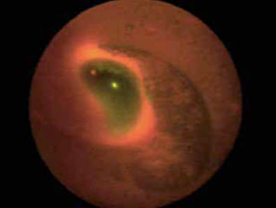
Scan of ear canal using Emission Reabsorption Laser Induced Fluorescence (ERLIF)
(Hidrovo and Hart)
The process referred to as Emission Reabsorption Laser Induced Fluorescence (ERLIF), and is based on a technology of emission and re-absorption. We Interestingly enough, we were developing this technology for measuring the thickness of oil films in internal combustion engines as they ran - a very different application from ear canals. Basically, the technology consists of taking a fluid, illuminating it with light, and putting two different dyes inside that fluid. The light, as it passes through the fluid, gets absorbed by both dyes and fluoresces back. From there, the dye is selected such that one fluoresces and transfers back through the liquid with almost no interruption, and the other one fluoresces and gets reabsorbed, in part, by the other dye. The result is that when you image these two different frequencies of light coming back, you can ratio them and they give an extremely accurate measurement of the liquid thickness.
We then created an expanding membrane, which would hold the liquid with the dyes, and be inserted in the ear canal. With the liquid and dyes inside the membrane, we can image it with a device that looks very similar to a fiberscope. Using just a few images, we can take a 3D image of the ear canal. So, it's an inexpensive, robust system.
Smaka: How would your scanner differ from current impression scanners currently available to audiologists?
Hart: We are obtaining a direct in-ear measurement of the ear canal, without the error-prone intermediary of the impression material;no matter how good a desktop scanner is, the result is limited by the inaccuracies in the impression material. In addition, they do not measure ear canal compliance, which is a key data point for hearing aid manufacturers.
As for the current desktop impression scanners, I'm not an expert but my understanding is that they either use a profilometer-type system, where they measure the profile or rotation of the images, or they use a structured light-type system. The second type of system essentially uses the structure of the light that it receives back onto a camera to resolve three dimensions. They can be reasonably accurate, but there are some limitations with these systems for measuring the internal ear. One issue is that skin is translucent and there is earwax, hair, and so forth inside the ear canal that these systems do not account for. The other issue is that they do not measure the compliance of the ear canal, which I think is a major component for fitting hearing aids. I believe, once we get more data, people will find that the compliance of the ear canal is a big factor in designing the fit of a hearing aid, getting it to feel comfortable and ensuring an appropriate seal of the hearing instrument or earmold.
Smaka: Do you have any projections what something like this could cost in an audiology practice?
Hart: I would hate to guess at this point. We have proven that it will work, but it is not yet ready for the clinic. By the time it goes from a laboratory environment to a system that an audiologist would use, it's hard for me to project. Fundamentally, from an engineering perspective, it is a very simple device. There is nothing complex in the optical system and the software and hardware are very straightforward. My feeling is that as 3D imaging devices go, it certainly falls well below the average in terms of cost. My guess is that it would be in the price range of current impression scanners. We've been looking at the market in general and what the needs are, and I just don't believe there's room in the market for a particularly expensive device. I think if anybody is going to develop a useful device, it has to be on the order of the equipment that audiologists buy now, and it can't be something outrageously expensive.
Smaka: If audiologists did not have to purchase impression material, and had less remakes for fit issues, there would be cost savings for the professional as well.
Hart: That's certainly the hope. You eliminate the impression material, and you also eliminate the trouble of having to have another step. You do not have to have an impression scanner sitting in the office. For audiologists who are still mailing ear impressions to manufacturers and earmold labs, this would eliminate this step and the associated delays. By eliminating the hardware, the materials, and the processing, we hope it would be a very pleasant experience for both the audiologist and the patient to use this system.
I'm not an audiologist, but the current system always struck me as a little intimidating for a patient, to have a professional approaching your ear with what looks like a caulking gun, or a syringe.
Frankly, with this new scanning technique, it's also fun to see the three dimensions evolve on the screen in real time. It's a very quick process;you can see right as you look on the screen the three-dimensional image forming. This turned out to be a huge selling point for patients when we did this with teeth in the dental industry. Patients became very comfortable feeling that this was a high-tech process, and they developed a great deal of confidence that what they were getting was something significant and not something arcane.
Smaka: That is an excellent point. We see that with video otoscopy, real ear measurements, hearing aid programming and other clinical techniques. When patients are more involved in the process, they tend to feel more confident in the outcome. Do you see other applications for your technology besides audiology, for example, for ear surgeons or other related fields?
Hart: Certainly. We have talked with ENTs, and they said they would love to be able to take scans for work they do, and there are also military applications, hearing suppression devices, and communication devices.
Smaka: What are the next steps? When do you expect that this system would be ready for clinical use?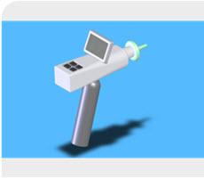
Ear canal scanner - artist's impression
Hart: We are moving forward with commercializing this technology. This research was sponsored by a group called the Deshpande Center here at MIT, which is a center specifically geared toward taking technology that would be useful to the public in short-term, and spinning it out into companies that can bring this to the market. Therefore, we are currently looking at spinning the technology out of MIT and forming a company. Our hope is that we'll either spin this out into a company or we will sell it off to a company able to commercialize it in short order. Either way, my guess is probably a year or two before it will be in common use.
Smaka: Would you be interested in having audiologists field-trial or provide clinical input for your device?
Hart: We are actually very much interested in input. We'd very much like to hear suggestions on features that people would enjoy or would like to see in our device. For instance, just recently we started talking with audiologists who were telling us that one of the things they struggle with are all the forms that they have to fill out to order hearing aids, and how it would be really great if we could integrate all that into our system. Then, not only could you scan the ear, but you could also fill the order form out online and send it out all through one compact system, without going through 50 different areas. We originally had not thought about integrating those two ideas together, and this was a useful suggestion. We're currently learning about audiologists' workflow, what kind of problems they run into during their clinical activities, and how our device might solve these problems. We are very new to this area;therefore, we are very appreciative of anything we can learn from professionals in the field.
Smaka: Where can people contact you if they did want to provide input?
Hart: The easiest thing is to email me at [email protected].
Smaka: Dr. Hart, thank you for speaking with us about this emerging technology.
Hart: Thank you for your interest, Carolyn.
Dr. Hart can be contacted at [email protected].
