
Auditory electrophysiological assessment of patients and research subjects is well established in clinical practice and dates back to the early 1930's when electroencephalographic (EEG) measurement was introduced to medicine. In the early days, measurements were restricted to the higher amplitude cortical potentials, and the much smaller brainstem potentials awaited discovery of the signal averaging computer which gained medical application in the 1960's. In the second half of that decade there emerged reports of measuring VIIIth cranial nerve and auditory brainstem evoked potentials that were stunningly microscopic. These electrical events were on the order of a few tenths of a microvolt whereas earlier recorded cortical potentials were hundreds of microvolts. By coupling the signal averaging computer to established recording methods of the time, these much smaller potentials could be identified. Also, precisely manipulating a capture 'window' from several hundred milliseconds in duration down to 10 milliseconds or less enabled targeting the earliest occurring electricity in the auditory system at the inner ear and the few milliseconds following that electrical genesis. Clearly, this was a monumental break-through in medicine.
What happened to the discipline of audiology following discovery of the auditory brainstem response (ABR) is well understood by most practicing clinicians. In its initial application it was determined to be sensitive to retro-cochlear pathology, and for the first time in the history of the profession 'sensory neural hearing loss' could be more accurately defined as either 'sensory' or 'neural'. The ABR test was found to be more sensitive to acoustic neuroma (vestibular schwannoma) than acoustic reflex measurement and all retrocochlear behavioral test procedures combined. The test identified the presence of a small acoustic neuroma when computerized tomography (CT) scanning could not. Much has happened in the intertwined disciplines of electrophysiology and radiology since the 1970's, but one characteristic of each discipline is noteworthy: First, radiology has progressed from producing an out of focus black and white smear to a detailed photographic quality magnetic resonance image (MRI) while second, the ABR test has become user-friendly. To be sure, the mission of research and development (R&D) was distinct for each discipline. Radiology set out to achieve better imaging and improved diagnostic capability of test procedures which was accomplished. During the same time period ABR instrumentation progressed from toggle switches and racks of components to windows-based software without appreciable improvement in response 'imaging'. The challenge for auditory electrophysiologists is to improve test sensitivity through manipulation of available parameters (signals, filters, gain, time base) with an effort to keep the test viable clinically and not replaced entirely by MRI, as has occurred in many clinics. It should be well understood by medical practitioners that ABR and MRI represent very different topic areas. The ABR falls under the category of 'physiology', or how the system works, and the MRI is strictly 'anatomical', or how the system appears. Responsible medicine demands careful assessment of the patient's anatomical as well as the physiological function for diagnostic accuracy. The radiologist is aided immensely by responsible R&D. The audiologist must compensate for an R&D deficit with clever manipulation of test parameters that have remained largely unchanged for decades. For example, many clinics now use a rapid stimulus rate to help identify possible compromise to the auditory nervous system through a process of neural adaptation or 'fatigue'. Another example of clever manipulation of ABR test parameters is the Vestibular Evoked Myogenic Potential (VEMP) test.
A Quick Guide to VEMP Testing
Description of the Vestibular Evoked Myogenic Potential (VEMP) Procedure.
The VEMP procedure is an emerging diagnostic tool for identifying vestibular lesions. The VEMP procedure is non-invasive and causes little or no discomfort to the patient, unlike the caloric irrigation component of the videonystagmography (VNG) procedure. In addition, the test contributes an assessment dimension to the well-established VNG protocol. While caloric irrigation testing is focal to the lateral semicircular canal (LSC) and neural links to eye muscles, VEMP testing targets the vestibule and neural connections to the sternocleidomastoid muscles (SCM) of the neck. The VNG procedures evaluate semicircular canals and neural input to pons and cerebellar structures, while the VEMP procedure stimulates the saccule and connections through the medulla and into the neck. The procedures are complementary in that VEMP testing completes the lower brainstem and upper spinal column assessment component not targeted with VNG procedure. VEMP tests linear acceleration/gravitational orientation balance function, while VNG tests angular acceleration and central balance capability. Further, as noted, VEMP testing is tolerated well by the patient making it a useful adjunct procedure to VNG in a single test session.
Patients with true vertigo (hallucination of motion or the feeling of spinning), or a less specific feeling of imbalance or lightheadedness are routinely evaluated with a complete audiological evaluation and VNG testing. The most objective of the VNG subtests, the caloric irrigation procedure, grossly measures response of the LSC but does not assess the function of other vestibular sensory structures: namely, superior and posterior semicircular canals, saccule, and utricle (Akin and Murnane, 2001). Other procedures in the VNG sequence may identify posterior semicircular canal disorder (Dix-Hallpike Maneuver) or central lesion (direction changing nystagmus with positional change). However, often VNG findings are inconclusive or 'non-localizing' and do not always specify a locus of lesion. By including VEMP procedures in the clinical work-up of patients with imbalance, a more thorough assessment of the vestibular mechanism is accomplished, and in addition a specific vestibular sensory system is targeted.
The VEMP by definition is a short-latency electromyogram recorded from the tonically contracted SCM in response to high-intensity acoustic stimulation (Bickford et al., 1964; Cody and Bickford, 1969; Colebatch et al., 1994). With electrodes placed on the SCM muscles, waves occur at approximately 13-15 ms (p13 or wave I) and 21-24 ms (n23 or wave II) post stimulus delivery to the ear ipsilateral to the contracted SCM. The VEMP neural pathway consists of the saccule, inferior vestibular nerve, and vestibulospional tract (Colebatch and Halmagyi, 1992; Itoh et al., 2001; Murofushi et al., 1996a; Murofushi et al., 2001). The neural route of the VEMP is shown in Figure 1.
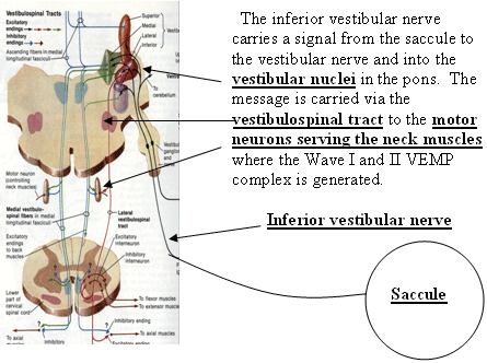
Figure 1. The neural route for the VEMP response.
More evidence of saccular stimulation is shown with experiments on guinea pigs and cats. Loud acoustic transients are shown to stimulate saccular afferents and are recorded electrophysiologically (Didier and Cazals, 1989; MuCue and Guinan, 1994; Murofushi et al., 1995, 1996b; Murofushi and Curthoys, 1997). Involvement of saccular tracts as well as the lower brainstem in normal hearing subjects are verified in additional reports (Townsend and Cody, 1971; Cazals et al., 1987; McCue and Guinan, 1994; Halmagyi and Colebatch, 1995; Robertson and Ireland, 1995; Colebatch et al., 1994; Itoh et al., 2001).
Clinical Applications
The VEMP gives important information about saccular and inferior vestibular nerve functions, which supplements caloric assessment of lateral semicircular canals, as well as other components of VNG procedure that target non-saccular vestibular systems. Recently, a mathematical calculation of VEMP amplitude was proposed (Young, Wu, Wu, 2002). Finding the difference in amplitude between the two sides divided by the sum of the amplitudes gives a ratio. When the ratio exceeds .36 the result indicates 'distended saccule' or saccular hydrops. This condition of excessive endolymph traditionally defines Meniere's Disease. This calculation is applied in all cases when the VEMP inter-aural VEMP amplitude differs notably. A case demonstrating this application is presented below. Response threshold is identified as the lowest signal intensity needed to acquire a repeatable waveform, which is 85-90 dB in normal subjects according to pilot data findings. Another recent discovery in the application of VEMP procedure clinically is identification of an inner ear pathology known as 'superior canal dehiscence' (SCD), or thinning or opening of the bony superior semicircular canal of the inner ear (Streubel, et al., 2001). Other studies report VEMP sensitivity to acoustic nerve tumor (Murofushi, et al., 2001), Meniere's Disease (Murofushi, et al., 2001; Shojaku, et al., 2001), brainstem stroke (Itoh, et al., 2001), and multiple sclerosis (Versino, et al., 2002).
It is reported that VEMP is mediated by an ipsilateral pathway. That is, VEMPs are recorded when the stimulus ear and contracted SCM are on the same side (Akin and Murnane, 2001; Halmagyi and Curthoys, 2000). This suggests that the auditory stimulation to the ear contralateral to the measured SCM muscle may not affect myogenic responses, and therefore that the presence of contralateral noise may not interfere with the recording of VEMP. However, a recent report by Takegoshi and Murofushi (2003) shows a change in VEMP with contralateral noise levels of 75 and 95 dB nHL. They studied 10 normal subjects and 10 patients with hemifacial palsy. Masking levels of 75 dB and 95 dB HL significantly (p<.05 reduced vemp amplitude. since the effect was greater in normal subjects than patients with hemifacial palsy they concluded amplitude reduction to be due stapedius muscle reflex that reduces conduction of stimulus inner ear. finding masking effects on is consistent clinical observations and also noted by gallaudet researchers ackley>
VEMP Test Protocol
Commercially available ABR instrumentation can be easily adapted to VEMP testing by changing all standard ABR parameters. The first and most obvious change is the location of electrodes on the patient. Active (non-inverting) electrode is placed on the superior aspect of the sternum with reference electrodes at each sternocleidomastoid muscle (Figure 2).
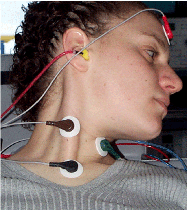
Figure 2. Electrode montage for VEMP involves placement on the sternum (non-inverting), and each sternocleidomastoid muscle with ground on the forehead. Test procedure requires contraction of each sternocleidomastoid by lifting the head and looking in the opposite direction of the test ear. This figure shows right ear stimulation and recording from the sternum - right sternocleidomastoid.
Acoustic signals require sufficient amplitude to disturb the otolithic membrane of the saccule. Low frequency tones (e.g. 500 Hz 2/0 Hanning Envelope) of 95 dBnHL provide reliable stimulation when delivered at a rate of less than 15/second (e.g. 5.1/sec.). Capture window needs to be adequate to record a wave that occurs around 25 ms after the stimulus (e.g. 50 ms window). Bandpass filtering must allow the low frequency response of myogenic activity (e.g. 10-2k Hz), and gain is reduced significantly from ABR requirements (e.g. 2k gain).
VEMP Response
The VEMP is recorded in the first 30 ms after the stimulus, with the first positive polarity peak occurring at 13-15 ms and the second negative polarity peak occurring at 22-25 ms (Figure 3). Response latency is somewhat dependent on stimulus parameters (frequency, duration, amplitude
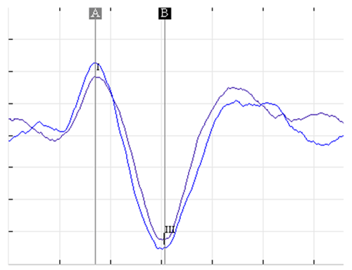
Figure 3. A normal VEMP response shows a positive polarity peak at about 13 ms and a negative polarity peak at 23 ms. Response latency varies with signal frequency, duration and to some extent with signal amplitude. The response is evident at 95 dB nHL.
VEMP Research
Studies conducted at the Gallaudet University Hearing and Balance Clinic show that the test is suitable for deaf and hard of hearing patients. Conductive hearing loss usually prevents VEMP responses, but cochlear damage seems to be independent of the VEMP waveform. Preliminary findings of Connexin Deafness and Waardenburg Syndrome suggest possible VEMP response abnormalities, and stimulus rate of 15-20/sec seems to degrade VEMP threshold. Future research will need to identify reliable test frequencies and signal durations other than the established norms for 500 Hz. Waveforms characterizing medulla pathology, inferior vestibular nerve pathology and saccular hydrops may be determined with detailed clinical studies.
ABR/VEMP Assessment of Patients with Imbalance
Clinical assessment of patients with symptoms of imbalance requires a battery of tests including videonystagmography (VNG), audiometric evaluation, ABR and VEMP as a minimum initial work-up. Electrocochleography (ECochG), Glycerol Testing and MRI may follow. A recommended first test protocol is the ABR/VEMP battery. The ABR is recommended to help identify the small percentage of patients who experience imbalance secondary to tumor. The VEMP helps to identify saccular, inferior vestibular nerve branch, and medulla pathology. The combined protocol requires about 45 minutes of test time and causes little or no discomfort to the patient.
Case Studies
Case 107. This is a 43 year-old profoundly deaf subject who was referred to the Balance Center because of complaint of vertigo when she hears loud sound, or Tullio phenomenon. The patient discovered that with her right hearing aid adjusted to full volume with loud noise in the environment, she became vertiginous. This was verified in the Balance Center after attempting to induce vertigo with the left hearing aid and failing. With right hearing aid on but without noise in the environment, similarly no vertigo was induced. However, after 45 seconds of environmental noise including multi-talker loud conversation and music and amplified through her right hearing aid, vertigo resulted and was documented as nystagmus (rapid eye movements) with videonystagmography. VEMP testing was performed (Figure 4) with the following results:
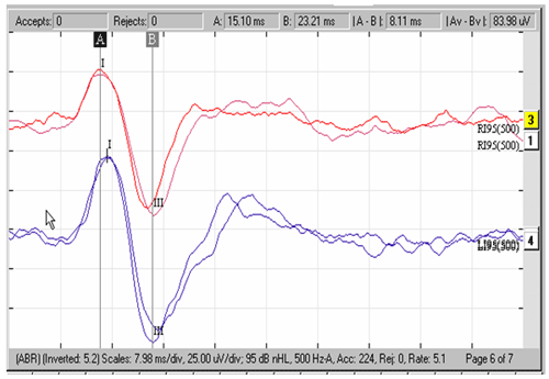
Figure 4. VEMP tracings for Case 107.
This VEMP tracing for right (top) and left (bottom) shows a symmetrical response with respect to wave latency (time delay of waves), but shows a reduced amplitude for the right side. Response amplitude for the right is 80 uV and for the left is 110 uV suggesting a disorder of the right saccule. Calculation of the 'augmented VEMP' (LA - SA/R + L>.36; where 'LA" and 'SA' are large and small amplitudes and 'R' and 'L' are 'right' and 'left' amplitudes) is not significant for saccular hydrops (Young, Wu & Wu, 2002). Further VEMP testing discovered reduced VEMP threshold which may be consistent with SCD, but CT scans of the superior and posterior semicircular canals were negative for SCD.
Case 117. This patient is a 45 year-old male who has been deaf since birth and from a deaf family. He has Connexin 26 and 30 deafness, according to genetic studies conducted at the Gallaudet Genetics Clinic. He was diagnosed in 1976 with Meniere's disease, and he was treated several months later with migraine medication. He continued to take the medication until recently when he stopped taking the meds because of rapid and irregular heart rate. He became vertiginous again and was gradually reintroduced to a low dose of his original migraine medication. He was seen for VEMP testing at the Gallaudet Balance Center.
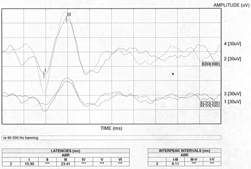
Figure 5. Right side VEMP tracings for Case 117.

Figure 6. Left side VEMP tracings for Case 117.
Figures 5 and 6 shows the VEMP results for a 45 year-old deaf patient (case # 117) showing ipsilateral (upper tracing) and contralateral (lower tracing) records for right and left sides. Wave latencies are within normal limits bilaterally (lower grid). Amplitude for the right side (127 uV) is normal, but left side amplitude (39 uV) is significantly reduced giving an augmented VEMP calculation probable site of 127-39/127+39 = 0.53 lesion, which is significant for saccular hydrops should assist in this patient's ongoing otoneurologic treatment. ENG caloric test results showed a 21% left reduced vestibular response which is marginally indicative of left side damage also. Follow-up evaluation by an otoneurologist is ongoing. Tracings are 'reversed' because of active (non-inverting) electrode placement on the sternum and referencing each SCM muscle.
EMG Monitoring
A quick note on EMG monitoring. Contraction of the sternocleidomastoid muscle is recommended, although the above cases do not describe this aspect of the procedure. Commercially available evoked potential test instruments now include electromyographic monitoring of SCM contraction strength during testing. Because the amplitude of the VEMP response is affected by SCM contraction strength, ongoing EMG monitoring of SCM contraction may improve amplitude repeatability of the waveform.
References
Ackley, R.S., Tamaki, C. (2003). Application of VEMP to Deaf and HoH Patients: A Clinical Study. American Speech-Language and Hearing Association National Convention. Chicago, IL. November, 2003.
Ackley, R.S., Ferraro, J., Arenberg, I.K. (1994), Diagnosis of Patients with Perilymphatic Fistula, Seminars in Hearing, Vol. 15, No. 1, pp. 37-42
Akin, F.W., Murnane, O.D., 2001. Vestibular evoked myogenic potentials: preliminary report. J. Am. Acad. Audiol. 12, 445-452.
Akin, F.W., Murnane, O.D., 2004. Vestibular evoked myogenic potentials. Insights in Practice: Clinical Topics in Otoneurology. GN Otometrics. April, 2004.
Aran, J.M., Pajor, A.M., de Sauvage, R.C., Erre, J.P., 2000. Role of the efferent medial olivocochlear system in contralateral masking and binaural interactions: an electrophysiological study in guinea pigs. Audiology 39(6), 311-21.
Bickford, R.G., Jacobson, J.L., Cody, D.T.R., 1964. Nature of averaged evoked potentials to sound and other stimuli in man. Ann. N.Y. Acad. Sci. 112, 204-223.
Cazals, Y., Erre, J.P., Aurousseau, C., 1987. Eighth nerve auditory evoked responses recorded at the base of the vestibular nucleus in the guinea pig. Hear. Res. 31, 93-98.
Cody, D.T.R., Bickford, R.G., 1969. Averaged evoked myogenic responses in normal man. Laryngoscope 79, 400-416.
Colebatch, J.G., Halmagyi, G.M., 1992. Vestibular evoked potentials in human neck muscles before and after unilateral vestibular deafferentation. Neurology 42, 1635-1636.
Colebatch, J.G., Halmagyi, G.M., Skuse, N.F., 1994. Myogenic potentials generated by a click-evoked vestibulocollic reflex. J. Neurol. Neurosurg. Psychiatry 57, 190-197.
Didier, A., Cazals, Y., 1989. Acoustic responses recorded from the saccular bundle on the eighth nerve of the guinea pig. Hear. Res. 37, 123-128.
Ferber-Viart, C., Dubreuil, C., Duclaux, R., 1999. Vestibular evoked myogenic potentials in humans: a review. Acta Otolaryngol. 119, 6-15.
Halmagyi, G.M., Colebatch, J.G., 1995. Vestibular evoked myogenic potentials in the sternomastoid muscle are not of lateral canal origin. Acta Otolaryngol. Suppl. (Stockholm) 520, 1-3.
Halmagyi, G.M., Curthoys, I., 2000. Otolith function tests. In: Herdman, S.J. (Ed). Vestibular Rehabilitation. Philadelphia: FA Davis, 196-214.
Itoh, A., Kim, Y.S., Yoshioka, K., Kanaya, M., Enomoto, H., Hiraiwa, F., Mizuno, M., 2001. Clinical study of vestibular-evoked myogenic potentials and auditory brainstem responses in patients with brainstem lesions. Acta Otolaryngol. Suppl. 545, 116-119.
McCue, M.P., Guinan, J.J. Jr., 1994. Acoustically responsive fibers in the vestibular potentials in the vestibular nerve of the cat. J. Neurosci. 14, 6058-6070.
Murofushi, T., Curthoys, I.S., 1997. Physiological and anatomical study of click-sensitive primary vestibular afferents in the guinea pig. Acta Otolaryngol. (Stockholm) 117, 66-72.
Murofushi, T., Curthoys, I.S., Topple, A.N., Colebatch, J.G., Halmagyi, G.M., 1995. Responses of guinea pig primary vestibular neurons to clicks. Exp. Brain Res. 103, 174-178.
Murofushi, T., Halmagyi, G.M., Yavor, R.A., Colebatch, J.G., 1996a. Absent vestibular evoked myogenic potentials in vestibular neurolabyrinthitis: an indicator of inferior vestibular nerve involvement? Arch. Otolaryngol. Head Neck Surg. 122, 845-848.
Murofushi, T., Curthoys, I.S., Gilchrist, D.P., 1996b. Responses of guinea pig vestibular nucleus neurons to clicks. Exp. Brain Res., 111, 149-152.
Murofushi, T., Shimizu, K., Takegoshi, H., Cheng, P.-W., 2001. Diagnostic value of prolonged latencies in the vestibular evoked myogenic potential. Arch. Otolorngol. Head Neck Surg. 127, 1069-1072.
Ochi, K., Ohashi, T., Nishino, H., 1999. Variance of vestibular-evoked myogenic potentials. Laryngoscope 111, 522-527.
Polyakov, A., Pratt, H., 2003. The cumulative effect of high click rate on monaural and binaural processing in the human auditory brainstem. Clin. Neurophysiology 114(2), 366-75.
Robertson, D,D., Ireland, D.J., 1995. Vestibular evoked myogenic potentials. J. Otolaryngol. 24, 3-8.
Shojaku, H., Takemori, S., Kobayashi, K., Watanabe, Y., 2001. Clinical usefulness of glycerol vestibular-evoked myogenic potentials: preliminary report. Acta Otolaryngol Suppl 545:65-8.
Streubel, S.O., Cremer, P.D., Carey, J.P., Weg, N., Minor, L.B., 2001. Vestibular-evoked myogenic potentials in the diagnosis of superior canal dehiscence syndrome. Acta Otolaryngol Suppl 2001;545:41-9.
Tamaki, C., Ackley, R.S., 2003. Vestibular Evoked Myogenic Potentials: Contralateral Masking Effects. Hearing Research. In Review.
Takegoshi, H., Murofushi, T., 2003. Effect of white noise on vestibular evoked myogenic potentials. Hear. Res. 176, 59-64.
Townsend, G.L., Cody, D.T.R., 1971. The averaged inion response evoked by acoustic stimulation: its relationship to the saccule. Ann. Otol. Rhinol. Laryngol. 80, 121-131.
Tsutsumi, T., Komatsuzaki, A., Noguchi, Y., Tokano, H., Kitamura, K., 2001. Postoperative vestibular-evoked myogenic potentials in cases with vestibular schwannomas. Acta. Otolaryngol. 121, 490-493.
Ungan, P., Yagcioglu, S., 2002. Origin of the binaural interaction component in wave P4 of the short-latency auditory evoked potentials in the cat: evaluation of serial depth recording from the brainstem. Hear. Res. 167(1-2), 81-101.
Wu, C.H., Murofushi, T., 1999. The effect of click repetition rate on vestibular evoked myogenic potential. Acta Otolaryngol. (Stockholm) 119, 29-32.
Young, Wu, C.C., Wu, C.H. (2002) Augmentation of vestibular evoked myogenic potentials: an indication for distended saccular hydrops. Laryngoscope. 112 (3). 509-512.
