
During electronystagmography (ENG) or videonystagmography (VNG), the examiner often faces a critical decision. Do the test results accurately reflect the physiologic and pathologic status of the patient or do they represent artifacts caused by technical errors? The answer is not always simple and wrong decisions can have serious consequences. For example, artifacts can cause the examiner to either miss an abnormal finding or misidentify a normal result as abnormal.
To identify technical errors, the examiner must have a basic knowledge of the capabilities and limitations of the ENG or VNG equipment. Also, the examiner must be familiar with the underlying physiology of the vestibular function tests. Technical errors must be suspected when results can not be explained by any known physiology or pathology of the vestibular and oculomotor pathways.
Although technical errors in ENG and VNG can be due to various causes, there are a few errors that are more common than others. In this article, I will describe how to identify some of these common errors and ways to avoid them.
1. Failure to perform physical examination of eye movements
Examiners must perform a thorough examination of the eye movements before the actual ENG or VNG testing. Failure to do so, can cause a number of different errors in different subtests.
First, the physical examination can reveal if the eyes movements are conjugate. With the standard electrode arrangement in ENG, horizontal movements of the right and left eyes are averaged based on the assumption that both eyes move identically. Similarly, most VNG systems also average the movements detected by the right and left cameras. In VNG, both horizontal and vertical eye movements undergo the averaging process. Failure to recognize disconjugate eye movements will result in faulty measurement of eye movements throughout the test because averaged signals do not accurately reflect actual movements of either eye.
Depending on the type of disconjugate eye movements, the examiner must change the standard protocol to record either movements of one eye only or to record movements of both eyes independently. An example of a case where eye movements must be recorded from one eye only, includes a patient who has significant reduction in the range of motion of one eye. This is usually caused by misalignment of the eyes (strabismus). Another example is a patient with one prosthetic eye. To record from one eye only in ENG, the examiner should move the electrode from the temple on the side of the affected eye to the bridge of the nose and place the vertical electrodes around the unaffected eye (Figure 1A). In VNG, the procedure for recording from one eye only, depends on the manufacturer of the equipment. Some VNG systems allow monocular recording. In others, one camera may have to be turned off manually by decreasing the brightness or other similar procedures.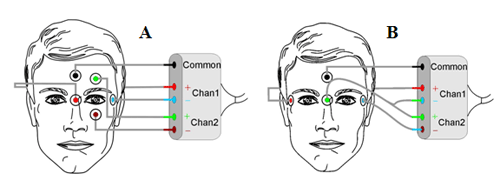
Figure 1. A) Electrode arrangement to record from one (left) eye only. B) Electrode arrangement to record horizontal movements of each eye independently.
In some cases, the range of motion is identical for both eyes but velocities differ during eye movements. For example, in patients with internuclear ophthalmoplegia, either one or both eyes are significantly slower during adduction (movement toward the nose) compared to the other eye. In this case, movements of both eyes must be recorded to document the difference between the eye velocities. In 4-channel ENG systems, the electrode arrangement in Figure 1A for the left eye can be duplicated for the right eye so that both horizontal and vertical movements can be recorded independently. However, most ENG systems are limited to two channels. In that case, vertical eye movements have to be sacrificed so that the horizontal eye movements can be recorded independently. This can be done by placing an electrode on the bridge of the nose and changing the electrode connections according to Figure 1B. Most VNG systems are capable of independent recording of right and left eye movements but the default setting, which is usually set to average right and left eye movements, may have be changed. Consult the manufacturer's manual for the exact procedure.
The second reason a thorough examination of eye movements can reduce technical errors is that it can reveal small eye movements. During the physical examination, the examiner observes the patient's eye movements in five primary gaze positions (center, 30 degrees right, 30 degrees left, 30 degrees up, and 30 degrees down). An experienced examiner can detect nystagmus as small as 0.5 degree/sec. ENG can theoretically measure nystagmus with the intensity of about 1 degree/sec. In practice however, the nystagmus intensity has to be greater than 2-3 degrees/sec before it can be differentiated from noise. Therefore, the examiner must rely on the result of the physical examination to determine if the eye movement tracings depict actual eye movements or they are noise. Figure 2 demonstrates the difficulty in identifying small nystagmus (about 2-3 degrees/sec) from the tracings although the nystagmus can be seen clearly in the accompanying video. This issue is obviously more important in ENG because VNG tracings are obtained by tracking the pupils. Nonetheless, physical examinations are still valuable adjuncts to VNG because they provide early warning as to which parts of the test require video recordings to document small eye movements.
Finally, a physical examination of eye movements can prevent technical errors during saccade and tracking tests. The examiner can generate saccadic eye movements by holding up two fingers and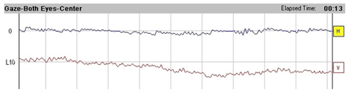
Figure 2. ENG tracings of the nystagmus seen in the corresponding video.
View Video Clip (Windows Media Video)
asking the patient to look back and forth between them. Tracking eye movements can be generated by asking the patient to follow the examiner's finger as it is moved slowly back and forth. Most common abnormalities such as ocular flutter, saccadic dysmetria, or abnormal tracking, can be observed during the physical examination. If the tracings suggest one of those abnormalities but the examiner can not confirm it from the physical inspection, a technical error should be suspected. For example, Figure 3 shows the results from the saccade test. The test seems to indicate abnormal accuracy (saccadic hypometria or undershoots). However, lack of confirming information from the physical examination should raise questions about the validity of the result. The underlying reason for the results in Figure 3 will be discussed next.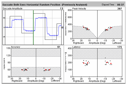
Figure 3. Invalid saccade test results caused by faulty calibrations.
2. Faulty calibrations
Calibration is the process of calculating a conversion factor so that the parameters measured by the recording device can be converted into eye movements. Therefore, a faulty calibration will cause the amplitude of the eye movements to be inaccurate in all of the subsequent subtests in the ENG or VNG test battery. Let us examine the calibration procedure in more detail and identify the causes that can lead to calibration errors.
Although the procedure is essentially the same, the underlying principles of calibration for ENG and VNG tests are completely different. In ENG, eye movements are determined indirectly by measuring the corneoretinal potential (CRP). The CRP is different from one person to another person and it changes over time in the same person. Therefore, calibration must be performed several times during the ENG test. Also, changes in the lighting conditions and vigorous movement of the patient can change the CRP and will require recalibration (Norman and Brown, 1999). In contrast, eye movements in VNG are determined directly by measuring the movements of the pupils. The calibration procedure in VNG is a simple conversion of the units from the camera coordinate system to the eye coordinate system. Therefore, recalibration is necessary only if the cameras or the goggles are moved with respect to the patient's eyes.
Historically, the calibration process consists of affixing fixation targets on the laboratory wall and asking the patient to look back and forth between the targets. The distance of the patient from the wall (typically around 4 feet) and the distance between the fixation targets are selected to generate eye movements with predetermined amplitude (typically plus or minus 10 degrees from the center gaze or total of 20 degrees). The examiner then adjusts the gain of the amplifier for the CRP until the pen deflections on the strip chart recorder correspond to predetermined amplitude (typically 20mm). The conversion factor between the eye movements and pen deflections is then calculated. For the typical eye movement amplitude of 20 degrees and the pen deflection of 20mm, the conversion factor is 1 degree of eye movement represented by 1mm of pen deflection. The procedure is performed for both horizontal and vertical channels.
The calibration procedure for the computerized ENG or VNG is similar to what is described above but there are a few differences. First, the fixation targets are generated by a light bar and the patient is instructed to follow the computer-generated dot. Some manufacturers use fast eye movements for calibration as the target jumps between fixation points. Others use slow eye movements as the target moves slowly between the same points. Second, most light bars are equipped with a range finder that determines the distance of the patient from the light bar. The computer alerts the examiner if the patient's distance from the light bar does not correspond to the recommended the distance. Finally, instead of allowing the examiner to manually adjust the gain, the computer automates this task. Most computerized ENG/VNG systems require the examiner to confirm when the eye and target movements match.
Faulty calibrations can originate from two types of errors: errors in determining the eye movements or errors in determining the target movements. One reason for error in determining eye movements is a tracing that is contaminated by noise or other artifacts. Another reason is a patient who looks around instead of following instructions and consistently fixating on the targets. In both cases, the examiner may be unable to accurately match the eye and target movements. In computerized ENG/VNG systems, this type of error can be easily recognized if the examiner allows the calibration process to continue for a few more seconds after confirming the match between the eye and target movements. Unfortunately, many examiners terminate the calibration process and miss the opportunity to recognize the mismatch between the eye and target movements. Even in those cases, the examiner can still recognize this type of error during the oculomotor tests. The saccade results in Figure 3 appear to be abnormal showing saccadic undershoot and slowing. However, an inspection of the eye and target movements shows that the amplitude of eye movements is significantly less than the amplitude of target movements. That is physiologically impossible because the eyes must always begin and end on the target regardless of what happens in between. If this were a true case of saccadic undershoot, the patient would have made multiple attempts and eventually would have reached the target. In other words, the eye position tracing would have looked like a staircase. Similarly, this type of error can be recognized in the tracking test if the peaks of eye and target movements do not match. Again, in the case of a true tracking abnormality, the patient will make multiple attempts and will not stop until reaching the target. Once this type of error is recognized, calibration must be repeated. Some manufacturers allow the examiner to modify the conversion factor and reanalyze the tests results. For example in Figure 3, one can increase the gain until the eye and target amplitudes match and reanalyze the saccade test. This may save some time because the examiner can skip repeating the saccade test. It should be noted that this type of faulty calibration can not be corrected for tests such as caloric and position tests, where there are no target movements to be used as reference (Stockwell, 1994). For those tests, the amplitude of eye movements will be inaccurate.
Faulty calibrations can also be produced by errors in determining the target movement. Such errors are most likely related to the inaccuracy of measuring the distance between the patient and the light bar (or the fixation targets). Consider a calibration setting in which the target movements are supposed to generate 20 degrees of eye deflection when the patient is set at 4 feet from the light bar. If the patient's actual distance from the light bar turns out to be 5 feet, after calibration, the amplitude of eye movements will be overestimated by 20%. Similarly, when the actual distance is 3 feet, the amplitude of eye movements after calibration will be underestimated by 32%. Unlike faulty calibrations caused by errors in determining the eye movements, there is no easy way of recognizing errors in the measurement of distance in the subsequent tests. The problem is confounded because some examiners do not seem to be aware of this error. For example, some choose to perform calibration during the position or caloric test while the patient is still in the supine position. The distance of the patient from the light bar can vary by as much as 2 feet-3 feet between sitting and supine positions. As a result, the amplitude of eye movements will be inaccurate and some abnormalities, such as abnormal positional nystagmus or hyperactive/ hypoactive caloric responses, can be misidentified. The only way to avoid this type of error is to make sure that the distance of the patient from the light bar is accurate before each calibration.
How often should the calibration procedure be repeated throughout the test to minimize calibration-related errors? As discussed earlier, calibration does not have to be repeated in VNG unless the goggles are moved. In ENG, changes in the lighting conditions and rapid changes of the patient's position always affect the CRP and require recalibration. Changes in the lighting conditions can be minimized if the entire test is performed in a dimly-lit environment. Even without those changes, CRP varies over time. It is estimated that the maximum change in the CRP occurs after 7-12 minutes of dark adaptation and 6-9 minutes after exposure to light (Lightfoot, 2004). Therefore, it is logical to repeat the calibration about every 10 minutes. In the most common ENG protocols, calibrations are performed at the beginning, following the oculomotor test, and following the position test. Recalibrations for the caloric test require further discussion. Most standards, such as those proposed by CHABA (1992) and ANSI (1999), call for recalibration before each irrigation. This is sound advice for non-computerized ENG. In those systems, recalibration means verifying the current calibration and making usually minor adjustments, when necessary. In computerized ENG, recalibration requires discarding the existing calibration and performing an entirely new calibration. Norman and Brown (1999) have demonstrated an average change of 17% between two calibrations even though the baseline CRP did not change significantly during caloric testing under constant lighting conditions. This suggests that in computerized ENG, calibration before each caloric irrigation can actually increase the chance of producing calibration errors. This problem could have been avoided if the computerized ENG systems provided a procedure for verifying the calibration just as in non-computerized ENG devices. Unfortunately, such a procedure is not currently available. There are at least two ways of overcoming this limitation. First, the examiner can use a mock saccade or tracking test before each irrigation to verify the calibration. When the target and eye amplitudes differ significantly, recalibration is warranted. Otherwise, the examiner can proceed with the irrigation. The examiner can also place fixation dots on the ceiling and ask the patient to look back and forth between them before each irrigation. Another mock test, such as position test, can be used to record the resulting eye movements. Again, recalibration is unnecessary unless the eye movement amplitudes change significantly from one test to another. The latter procedure also avoids the cumbersome process of sitting the patient up for calibration before each irrigation.
3. Failure to maintain a steady level of alertness
In the ENG or VNG test battery, the examiner must alert the patient during tests performed in the absence of fixation (Barber and Stockwell, 1980). In particular, during position and caloric tests, failure to maintain a steady level of alertness will result in the nystagmus appearing and disappearing intermittently based on the level of alertness. In some cases, the nystagmus may be missed completely without adequate alerting. If the loss of alertness occurs during the strongest part of the caloric response, the peak nystagmus velocity for that irrigation will be miscalculated.
To avoid these problems, the examiner should be prepared to ask the patient questions at a steady pace. Most patients do not require vigorous tasking but some are hard to alert. The exact nature of questions is less important as long as they are moderately challenging to the patient. The examiner should have a prepared list of questions with different levels of difficulty. If the nystagmus continues to be intermittent, the examiner should switch to a different set of questions. Once the patient's interest and knowledge level are established, the examiner should keep a steady pace and avoid long pauses.
4. Failure to elicit physiologically-valid caloric responses
The main assumption in the caloric test is that all four irrigations provide equally strong stimulation to the labyrinth. This assumption depends on a number of factors. Some can be directly controlled by the examiner. Examples of factors that can be controlled by the examiner are temperature, volume, and duration of irrigation. In addition, the examiner must make sure that there is no cerumen blocking the tympanic membrane and must maintain a constant level of alerting throughout the test. Some factors, such as the ear anatomy, body temperature, or perforations, are not controllable. The examiner must make a note of any factor that can affect the validity of the caloric test.
Sometimes caloric responses are invalid despite the examiner's best effort. Usually, invalid results can not be explained by any known physiology or pathology of the vestibular system. The set of physiologically-valid caloric responses is relatively small. In one set, all four caloric responses are approximately equal. This type of response is generated in patients with normal vestibular function and in those with bilaterally hypoactive or hyperactive function. In another valid caloric test, warm and cool irrigations generate approximately equal responses from each ear but the total response from the right ear is significantly different from the total response from the left ear. This type of response is generated in patients with unilaterally reduced vestibular function. Sometimes, the total responses from the right and left ears are approximately equal but nystagmus beating in one direction is significantly stronger than nystagmus beating in the opposite direction. This represents directional preponderance. There are two types of directional preponderance. The first type is caused by the presence of preexisting nystagmus in the supine position. In this case, caloric responses are equally strong in both directions, but the baseline or the starting point of the caloric response is shifted by an amount equal to the slow phase velocity of the pre-existing nystagmus. As a result, peak nystagmus intensities in the direction of the shift are higher than peak nystagmus intensities in the other direction. The second type of directional preponderance is characterized by caloric responses that are truly stronger in one direction without any baseline shift. This type is called gain asymmetry and was first identified by Halmagyi, Cremer, Anderson, Murofushi & Curthoys (2000). Directional preponderance due to baseline shift is common but gain asymmetry is extremely rare and is seen in only 1% of patients. Any combination of the physiologically-valid sets described here also constitutes a valid caloric test.
Invalid caloric responses can result from a number of problems. In most computerized ENG and VNG systems, the software determines the peak caloric responses by analyzing the nystagmus. Even the most sophisticated algorithms occasionally fail to correctly identify the peak responses. If the system allows manual inspection and cleaning of tracings, the examiner should do so for all caloric irrigations. If the system does not allow intervention in analyzing the response, the examiner should still estimate the peak response manually and compare it with the one calculated by the software. Remember that caloric responses always have the same shape regardless of their strength or direction. They usually start about 20 seconds after the onset of the irrigation (if the tympanic membrane is intact), rise to a peak about 30 seconds after the end of the irrigation (about 60-90 seconds after the onset of irrigation), and then decline to the baseline. If the caloric response does not look like that, the caloric test may be invalid. Other reasons for invalid caloric results are one or more faulty responses due to poor irrigations, lack of patient alertness, or bad calibrations.
Except for rare cases of gain asymmetry, the examiner can assess the validity of the caloric test by determining the baseline shift of the responses from each ear. In tests with questionable validity, these baselines shifts are significantly different. Figure 4 shows how the baseline shifts are calculated for the right and left ears by finding the midpoint of the response for each ear. In this case, the difference is over 16 degrees/sec (16 degrees/sec for the right ear, -0.5 degree/sec for the left ear) and more than likely represents invalid caloric results. Exactly how much of a difference constitutes a valid test, depends on the overall strength of the caloric responses. Smaller differences may signify an invalid test when the overall caloric responses are weak. Conversely, the difference between the baseline shifts has to be proportionally larger for the test to be considered invalid when the caloric responses are strong.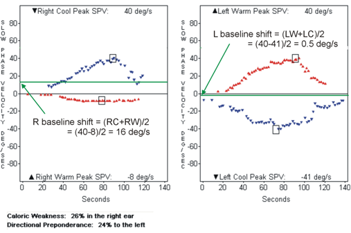
Figure 4. Checking the validity of the caloric results. One of the caloric responses is not as strong as the other three.
Figure 4 shows an example in which the response to one irrigation dominates the overall result. In this case, warm irrigation of the right ear produces significantly less intense response than the responses produced by the other three irrigations. This result can not be explained by any known physiology or pathology of the vestibular system and most likely represents a technical error. Possible causes for this error in the order of likelihood include: poor irrigation, faulty calibration, and loss of alertness. That is, the most likely scenario is that this result is generated by a patient who has normal vestibular function and all four caloric responses should be approximately equal. Therefore, the examiner should first repeat the right warm irrigation. The less likely scenario is that the patient has a unilateral vestibular dysfunction and the responses from the right ear should be significantly less than the responses from the left ear. Therefore, in the unlikely event that the peak caloric response does not change after repeating the right warm irrigation, the examiner should then repeat the right cool irrigation. Other scenarios are possible but they are highly unlikely. If after repeating two irrigations, the results still remain physiologically-invalid, the entire caloric test should be repeated at a later date. It is not recommended to perform more than six irrigations on the patient as a part of the same test.
Another example of a technical error in the caloric test is when the response to one irrigation is significantly greater than the responses produced by the other three irrigations. Possible causes for this error in the order of likelihood include: anxiety and hyper-alertness during the first caloric irrigation, faulty calibration, and poor irrigation (for the other irrigation of the same ear). Again, the examiner should first repeat the irrigation that produces significantly different response compared to the other three irrigations. If that does not resolve the technical error, the examiner should repeat the other irrigation in the same ear that produces the strong response.
Sometimes caloric responses for warm irrigations are significantly different than the responses to cool irrigations. This is called a temperature effect and represents a technical error. Fortunately the temperature effect does not affect calculations for unilateral weakness and directional preponderance. However, interpretation of the caloric test can become complicated when the temperature effect is combined with another technical error. Possible causes for temperature effect in the order of likelihood include: wrong temperature settings on the irrigator, faulty calibration, and significantly higher or lower than normal body temperature. Isolated cases of temperature effect do not cause major problems. However, if it is observed frequently, the examiner should ask the manufacturer of the irrigator to check the temperature setting. Also, it is a good idea to include the irrigator with other devices that undergo an annual calibration check.
Summary
Technical errors can and do happen during ENG and VNG testing. This article covers some but not all of the common errors. It is the responsibility of the examiner to prevent these errors when possible, and to detect and correct them when they occur.
References
American National Standards Institute (ANSI) (1999). Procedures for testing basic vestibular function. BSR S3.45-200X, revision of ANSI S3.45.
Barber, H.O., and Stockwell, C.W. (1980). Manual of Electronystagmography (pp. 142-152). St Louis: CV Mosby.
Committee on Hearing, Bioacoustics, and Biomechanics (CHABA) (1992). Evaluation of tests for vestibular function. Aviation, Space, and Environmental Medicine, 63 (Suppl. 2), A1-34.
Halmagyi, G.M., Cremer, P.D., Anderson, J., Murofushi, T., and Curthoys, I.S. (2000). Isolated directional preponderance of caloric nystagmus: I. Clinical significance. The American Journal of Otology, 21(4), 559-567.
Lightfoot, G.R. (2004). The origin of order effects in the results of the bi-thermal caloric test. International Journal of Audiology, 43(5), 276-282.
Norman, M., and Brown, E. (1999). Variations in calibration for computerized electronystagmography. British Journal of Audiology, 33(1), 1-7.
Stockwell, C.W. (2004, July). Three common errors in ENG testing. ENG Report, ICS Medical.
Common Errors in ENG/VNG
July 17, 2006
Continued and its subsidiaries provide professional education authored by qualified Subject Matter Experts for continuing education purposes. These materials are intended for educational purposes and do not constitute medical advice or a substitute for individual clinical judgment. Continued is not a clinical healthcare provider; the licensed professional is solely responsible for ensuring that the application of any techniques or information presented is within their legal scope of practice and jurisdictional requirements.

Related Courses
1
https://www.audiologyonline.com/audiology-ceus/course/guide-to-bithermal-caloric-testing-815
A Guide to Bithermal Caloric Testing
The purpose of this course is to provide an in-depth discussion of the caloric testing portion of the VNG/ENG test battery. Content will include descriptions of testing procedures, analysis and interpretation of results.
auditory, textual, visual
129
USD
Subscription
Unlimited COURSE Access for $129/year
OnlineOnly
AudiologyOnline
www.audiologyonline.com
A Guide to Bithermal Caloric Testing
The purpose of this course is to provide an in-depth discussion of the caloric testing portion of the VNG/ENG test battery. Content will include descriptions of testing procedures, analysis and interpretation of results.
815
Online
PT60M
A Guide to Bithermal Caloric Testing
Presented by Amanda Cerka Mroz, AuD, FAAA, CCC-A
Course: #815Level: Intermediate1 Hour
No CEUs/Hours Offered
The purpose of this course is to provide an in-depth discussion of the caloric testing portion of the VNG/ENG test battery. Content will include descriptions of testing procedures, analysis and interpretation of results.
2
https://www.audiologyonline.com/audiology-ceus/course/guide-to-bithermal-caloric-testing-36752
A Guide to Bithermal Caloric Testing
The purpose of this course is to provide an in-depth discussion of the caloric testing portion of the VNG/ENG test battery. Content will include descriptions of testing procedures, analysis and interpretation of results.
auditory, textual, visual
129
USD
Subscription
Unlimited COURSE Access for $129/year
OnlineOnly
AudiologyOnline
www.audiologyonline.com
A Guide to Bithermal Caloric Testing
The purpose of this course is to provide an in-depth discussion of the caloric testing portion of the VNG/ENG test battery. Content will include descriptions of testing procedures, analysis and interpretation of results.
36752
Online
PT60M
A Guide to Bithermal Caloric Testing
Presented by Amanda Cerka Mroz, AuD, FAAA, CCC-A
Course: #36752Level: Intermediate1 Hour
AAA/0.1 Intermediate; ACAud inc HAASA/1.0; AHIP/1.0; BAA/1.0; CAA/1.0; IACET/0.1; IHS/1.0; Kansas, LTS-S0035/1.0; NZAS/1.0; SAC/1.0
The purpose of this course is to provide an in-depth discussion of the caloric testing portion of the VNG/ENG test battery. Content will include descriptions of testing procedures, analysis and interpretation of results.
3
https://www.audiologyonline.com/audiology-ceus/course/using-eeg-biomarkers-to-understand-40915
The Use of EEG Biomarkers to Understand Cortical-Vestibular Interactions, in partnership with Vanderbilt University
This course examines the emerging evidence utilizing EEG biomarkers to gain a further understanding of cortical-vestibular interactions. Emphasis will be placed on understanding the history of EEG, clinical manifestations of cortical-vestibular dysfunction, and how EEG rhythms are currently being used in research to gain a better understanding of the vestibular system.
auditory, textual, visual
129
USD
Subscription
Unlimited COURSE Access for $129/year
OnlineOnly
AudiologyOnline
www.audiologyonline.com
The Use of EEG Biomarkers to Understand Cortical-Vestibular Interactions, in partnership with Vanderbilt University
This course examines the emerging evidence utilizing EEG biomarkers to gain a further understanding of cortical-vestibular interactions. Emphasis will be placed on understanding the history of EEG, clinical manifestations of cortical-vestibular dysfunction, and how EEG rhythms are currently being used in research to gain a better understanding of the vestibular system.
40915
Online
PT60M
The Use of EEG Biomarkers to Understand Cortical-Vestibular Interactions, in partnership with Vanderbilt University
Presented by Daniel Romero, AuD, PhD, Tricia Stanley, AuD
Course: #40915Level: Intermediate1 Hour
AAA/0.1 Intermediate; ACAud inc HAASA/1.0; ASHA/0.1 Intermediate, Professional; BAA/1.0; CAA/1.0; Calif. SLPAB/1.0; IACET/0.1; IHS/1.0; Kansas, LTS-S0035/1.0; NZAS/1.0; SAC/1.0; TX TDLR, #142/1.0 Non-manufacturer
This course examines the emerging evidence utilizing EEG biomarkers to gain a further understanding of cortical-vestibular interactions. Emphasis will be placed on understanding the history of EEG, clinical manifestations of cortical-vestibular dysfunction, and how EEG rhythms are currently being used in research to gain a better understanding of the vestibular system.
4
https://www.audiologyonline.com/audiology-ceus/course/addressing-fall-risk-pt-ot-38144
Addressing Fall Risk: PT, OT, and Audiology Assessment and Intervention, presented in partnership with Salus University
Multidisciplinary assessment and intervention of individuals at risk of falls is crucial in identifying functional and diagnostic factors as well as effective rehabilitation and prevention of future falls. This series identifies areas of collaboration and supportive information-sharing strategies between professions who commonly see individuals who fall and are likely to be injured due to a fall.
auditory, textual, visual
129
USD
Subscription
Unlimited COURSE Access for $129/year
OnlineOnly
AudiologyOnline
www.audiologyonline.com
Addressing Fall Risk: PT, OT, and Audiology Assessment and Intervention, presented in partnership with Salus University
Multidisciplinary assessment and intervention of individuals at risk of falls is crucial in identifying functional and diagnostic factors as well as effective rehabilitation and prevention of future falls. This series identifies areas of collaboration and supportive information-sharing strategies between professions who commonly see individuals who fall and are likely to be injured due to a fall.
38144
Online
PT180M
Addressing Fall Risk: PT, OT, and Audiology Assessment and Intervention, presented in partnership with Salus University
Presented by Bre Myers, AuD, PhD, Helena Esmonde, PT, DPT, NCS, Anna Grasso, OTD
Course: #38144Level: Intermediate3 Hours
AAA/0.3 Intermediate; ACAud inc HAASA/3.0; AHIP/3.0; ASHA/0.3 Intermediate, Professional; BAA/3.0; CAA/3.0; Calif. SLPAB/3.0; IACET/0.3; IHS/3.0; Kansas, LTS-S0035/3.0; NZAS/3.0; SAC/3.0; Tier 1 (ABA Certificants)/0.3
Multidisciplinary assessment and intervention of individuals at risk of falls is crucial in identifying functional and diagnostic factors as well as effective rehabilitation and prevention of future falls. This series identifies areas of collaboration and supportive information-sharing strategies between professions who commonly see individuals who fall and are likely to be injured due to a fall.
5
https://www.audiologyonline.com/audiology-ceus/course/contemporary-concepts-in-pediatric-vestibular-35586
Contemporary Concepts in Pediatric Vestibular Assessment and Management, presented in partnership with Seminars in Hearing
This 5-part webinar series is focused on the growing evidence of the need for pediatric vestibular evaluation, as well as the availability of successful treatment options for children. Guest editors, Dr. Devin McCaslin and Dr. Jennifer Christy along with a team of leading experts will present on select articles from a recent issue of the journal Seminars in Hearing (Issue 03 · Volume 39 · 2018).
auditory, textual, visual
129
USD
Subscription
Unlimited COURSE Access for $129/year
OnlineOnly
AudiologyOnline
www.audiologyonline.com
Contemporary Concepts in Pediatric Vestibular Assessment and Management, presented in partnership with Seminars in Hearing
This 5-part webinar series is focused on the growing evidence of the need for pediatric vestibular evaluation, as well as the availability of successful treatment options for children. Guest editors, Dr. Devin McCaslin and Dr. Jennifer Christy along with a team of leading experts will present on select articles from a recent issue of the journal Seminars in Hearing (Issue 03 · Volume 39 · 2018).
35586
Online
PT300M
Contemporary Concepts in Pediatric Vestibular Assessment and Management, presented in partnership with Seminars in Hearing
Presented by Sharon Cushing, MD, FRCSC, Katheryn Bachmann, PhD, Violette Lavender, AuD, Jennifer B. Christy, PhD, PT, Steven M. Doettl, AuD, PhD, Devin L. McCaslin, PhD, Kristen L. Janky, PhD, Amanda I. Rodriguez, PhD, AuD
Course: #35586Level: Intermediate5 Hours
AAA/0.5 Intermediate; ACAud inc HAASA/5.0; BAA/5.0; CAA/5.0; Calif. SLPAB/5.0; IACET/0.5; IHS/5.0; Kansas, LTS-S0035/5.0; NZAS/3.0; SAC/5.0
This 5-part webinar series is focused on the growing evidence of the need for pediatric vestibular evaluation, as well as the availability of successful treatment options for children. Guest editors, Dr. Devin McCaslin and Dr. Jennifer Christy along with a team of leading experts will present on select articles from a recent issue of the journal Seminars in Hearing (Issue 03 · Volume 39 · 2018).